J EMS Med Search

CLOSE


J EMS Med > Volume 2(1); 2023 > Article
Tan, Zarisfi, Tan, Sia, Ong, Ho, and Masuda: The role of passive leg raise during cardiopulmonary resuscitation in sudden cardiac arrest: a systematic review and meta-analysis

Abstract

Objective

Passive leg raise (PLR) is an experimental cardiopulmonary resuscitation (CPR) technique involving elevation of the lower limbs from the horizontal plane during CPR. This procedure is thought to improve clinical outcomes in cardiac arrest. By comparing the use of PLR to conventional CPR, we aimed to evaluate survival outcomes and secondary outcomes of neurologically intact survival and return of spontaneous circulation (ROSC).

Methods

In this systematic review, databases of PubMed, Embase, and Cochrane Library were searched for relevant studies from inception to May 1st, 2021. Studies were included if they reported the use of PLR during CPR. Random-effects meta-analysis was performed for the outcome of survival to 30 days.

Results

The search yielded 554 articles, of which six met the criteria for inclusion (three animal and three human studies). The most common implementation of PLR was elevation of the heels to a 20- to 45-degree angle at the hips. Two human studies reported higher survival at hospital admission for patients that underwent PLR, though the difference was not significant. Two human studies reported lower ROSC and neurologically intact survival, while two animal studies reported higher ROSC and neurologically intact survival. The pooled effect on survival to 30 days/survival to hospital discharge was not significant (P=0.68), although our analysis showed a trend favoring PLR-CPR.

Conclusion

Despite several animal studies showing benefit from PLR-CPR, there are no human data supporting its use in human cardiac arrest. Future research needs to ascertain the best positioning during CPR to optimize clinical outcomes.

INTRODUCTION

Sudden cardiac arrest (SCA) is a life-threatening medical condition highly-relevant to both out-of-hospital and inpatient settings. It involves the sudden cessation of cardiac activity where the victim becomes unresponsive with abnormal breathing and absence of circulation [1]. If active interventions are not started rapidly, SCA progresses to sudden cardiac death (SCD) [1]. Each year, SCD accounts for almost 25% of 17 million deaths worldwide, making it one of the most common causes of death [2]. Early commencement of effective cardiopulmonary resuscitation (CPR) improves survival [35]. Yet, clinical outcomes have remained poor, with a pooled global 30-day survival rate for out-of-hospital cardiac arrest (OHCA) patients of 10.7% [6]. Hence there remains an urgent need to discover therapeutics to improve clinical outcomes.
Passive leg raise (PLR) is an experimental CPR technique which involves elevation of the lower limbs from the horizontal plane during CPR [7]. It is a simple maneuver that works through intravenous volume expansion—where blood is rapidly shifted from the lower extremities towards the intrathoracic compartment to improve venous return [8]. Prior to 1992, it was recommended as part of the International Standards and Guidelines for CPR [9], but was subsequently removed due to paucity of supporting clinical evidence. Recent years have seen a resurgence of interest in this topic due to its ease of applicability and plausible physiological basis for improving cardiac output during CPR [8]. Various animal and human studies have suggested better neurological outcomes [7], as well as an improved cardiac preload and blood flow during chest compressions [10].
With renewed interest in the role of PLR in the treatment of SCA [11], there is a need to consolidate the literature, both preclinical and clinical, to clarify the role of PLR in SCA. In this systematic review and meta-analysis, we primarily hypothesized that PLR improves survival (30-day survival or survival to discharge) in SCA as compared to conventional (supine) CPR (C-CPR). We further hypothesized that PLR improves secondary outcomes, namely, neurologically intact survival and return of spontaneous circulation (ROSC). Clarity on the effectiveness of PLR in SCA will inform CPR techniques, translating to improved clinical outcomes.

METHODS

Literature search strategy

This systematic review was conducted in accordance with the Preferred Reporting Items for Systematic Reviews and Meta-Analyses (PRISMA) guidelines [12], and has been submitted to PROSPERO (ID: 281527). A comprehensive literature search was performed on PubMed, Embase and the Cochrane Library from inception to 1st May 2021. The search strategy was developed in consultation with a medical information specialist. The search strategy consisted of different combinations of the following search terms: leg declin*, PLR, leg elevat*, leg inclin*, leg ramp, leg rais*, passive leg rais*, resuscitation, CPR, cardiac arrest, CPR, cardiopulmonary resuscitation, and chest compress*. The search was performed by 2 independent reviewers (YKT & AFWH). During study screening for relevance, any disagreement was resolved by consensus with a senior author (MEHO). The reference lists of these articles were also screened, and hand-searched to identify further relevant studies.

Study and cohort selection

All studies (case reports, case series, preclinical studies, clinical trials and observational studies) that reported the use of PLR during CPR were included during the initial search. We subsequently excluded all studies that reported on other positions during CPR (such as head-up CPR), studies that did not have primary data, and those without an English translation.

Data extraction

Relevant quantitative data were extracted by 2 authors (YKT & AFWH) in the form of absolute counts and frequencies of events when appropriate. Any disagreement was resolved by consensus with a senior author (MEHO). Where available, the data included ROSC, survival to 30 days, survival to hospital discharge, neurologically intact survival, neurological outcomes, cerebral perfusion pressure, intracranial pressure, brain blood flow, coronary perfusion pressure (CPP), arterial blood gas results, right atrial pressure, and decompression aortic pressure.

Risk of bias assessment

The quality and risk of bias of included animal and human studies were assessed using the GRADE Assessment Tool [13]. The GRADE Assessment tool assesses quality of evidence in terms of study limitations, inconsistency, indirectness, imprecision and publication bias. These were graded with the consensus of 2 investigators (AFWH and YKT).

Statistical analysis

We presented the continuous variables as mean and standard deviation. Categorical variables were presented as frequencies and percentages. As there was substantial heterogeneity, we performed a random-effects meta-analysis on the odds ratio (OR) of survival to 30 days, comparing study arms randomized to PLR-CPR or C-CPR. Forest plots displayed individual and pooled ORs and 95% confidence intervals (95% CIs) for binary outcomes. Individual and pooled mean difference and 95% CI were presented for continuous outcomes. Two-tailed statistical significance was set at P<0.05. Between-study heterogeneity was assessed using the I2 statistic. Publication bias was assessed using funnel plots if there were 10 or more studies reporting the same outcome. All data analysis was conducted using the Cochrane Collaboration’s Review Manager (RevMan 5.4) Software Package.

RESULTS

Study selection and grading

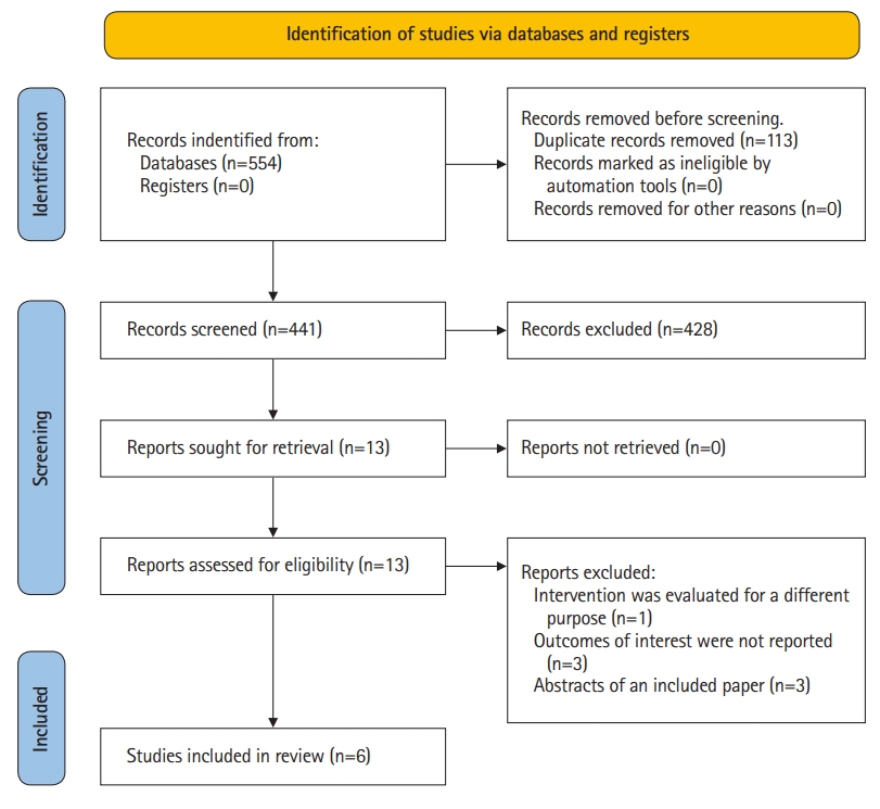
The search strategy yielded 554 studies, of which 113 studies were removed as duplicates. A further 428 studies were excluded after an initial screen of title and abstract as they did not report the use of PLR during CPR. Subsequently, 7 articles were excluded after full-text review. Finally, 6 eligible studies were included in our systematic review and meta-summary [7,8,1417]. The study selection process and reasons for excluding the 7 excluded studies were illustrated in the PRISMA-P 2020 Flow Diagram (Fig. 1).

Characteristics of included studies

Of the 6 studies included in the review, Zadini et al. [16], Dragoumanos et al. [7] and Qvigstad et al. [17] were experimental trials involving animal subjects, Holmen et al. [14] was an observational study involving human patients, and the remaining 2 were experimental human trials. Among the experimental human trials, Azeli et al. [15] was a randomized controlled trial (RCT) and Axelsson et al. [8] was a secondary analysis of a cluster trial. A meta-summary of included studies was presented in Tables 14 [7,8,1417].
Study design of human studies varied greatly. Axelsson et al. [8] was a secondary analysis of a cluster trial of mechanical CPR which aimed to assess partial pressure of end tidal carbon dioxide (pETCO2) levels that only included tracheal intubated patients with OHCA. The lone observational human study, Holmen et al. [14], retrospectively analysed OHCA cases over 3 years during which the emergency medical service (EMS) had implemented PLR within 5 minutes of CPR as part of their cardiac arrest protocol. Cases of non-compliance with the cardiac arrest protocol by the EMS study were then treated as the separate arm of the study. Meanwhile, Azeli et al. [15] randomized adult patients with OHCA to either PLR or C-CPR arms. However, details on the study arms and method of randomization were not reported.
For animal studies, Dragoumanos et al. [7] and Qvigstad et al. [17] were RCTs while the remaining study, Zadini et al. [16] was a single arm experimental trial. All animal studies involved porcine models of cardiac arrest where swines were subjected to a period of untreated ventricular fibrillation, which varied from 3 minutes to 8 minutes across study designs. However the aims across these 3 studies differed. Dragoumanos et al. [7] evaluated PLR’s effect on survival outcomes, hemodynamic performance and ROSC, while Qvigstad et al. [17] and Zadini et al. [16] solely evaluated hemodynamic performance and carotid blood flow respectively.

Experimental interventions and controls

The most common treatment among the 3 human studies was the bundling of PLR with an active compression-decompression device. Axelsson et al. [8] and Holmen et al. [14] implemented PLR as a 35 to 45 cm heel elevation which corresponded approximately to a 20- to 45-degree elevation at the hips. In contrast for the animal studies, Zadini et al. [16] implemented PLR as the Trendelenburg position where the pig was tilted 10, 20, and 30 degrees sequentially while Dragoumanos et al. [7] implemented PLR at 45 degrees (Table 2). All but one study used the supine position as the main control for comparison, as Zadini et al. [16] implementing a study design where each animal served as its own control. The remaining 5 studies had a proper control arm. However, amongst the 6 studies, Axelsson et al. [8], Holmen et al. [14] and Zadini et al. [16] and did not randomize their control arms, though it is worth noting that Holmen et al. [14] used propensity score matching to simulate balanced baseline prognostic factors.

Risk of bias

Quality of evidence was found to be high as evaluated by the GRADE framework in Table 5.

Study outcomes: primary and secondary outcomes

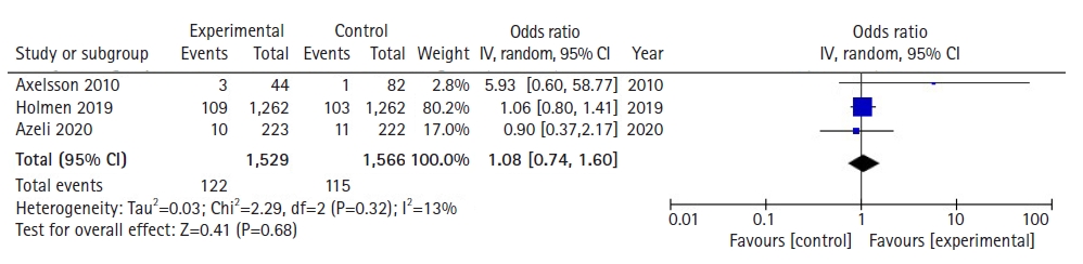
For the primary outcome of survival, 3 out of 6 studies included the outcomes of survival to 30 days/survival to discharge. These 3 studies all involved human patients. Meanwhile, only Axelsson et al. [8] and Azeli et al. [15] reported sufficient data for survival at hospital admission, and only Dragoumanos et al. [7] and Azeli et al. [15] reported on the outcome of neurologically intact survival. Dragoumanos et al. [7] was the sole study that accounted for the outcome of survival to 24 hours.
For the secondary outcomes, Axelsson et al. [8] and Dragoumanos et al. [7] detailed the outcome of ROSC. Similarly, Dragoumanos et al. [7] and Qvigstad et al. [17] reported on the outcome of CPP and Zadini et al. [16] was the sole study that investigated the impact of PLR on carotid blood flow.

Primary outcome: survival to hospital discharge/survival to 30 days

Azeli et al. [15] reported a lower proportion of PLR-CPR patients surviving, while both Axelsson et al. [8] and Holmen et al. [14] reported a higher proportion of patients that underwent PLR surviving at hospital discharge/to 30 days. However, all 3 comparisons were not statistically significant. It is also worth noting that Azeli et al. [15] reported survival to hospital discharge with cerebral performance category (CPC) 1 to 2 specifically.
With regards to survival to 30 days or survival to hospital discharge, a total of 3,095 patients across 3 studies were assessed based on pooled survival outcomes. Meta-analytic estimates for survival showed no statistically significant benefit for patients where PLR-CPR was conducted in comparison to patients that underwent C-CPR, as shown in Fig. 2 (OR, 1.08; 95% CI, 0.74−1.60; P=0.68). There was low heterogeneity (I2=13%).

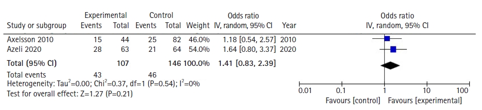
Primary outcome: survival at hospital admission

Only Axelsson et al. [8] and Azeli et al. [15] reported on the outcome of survival at hospital admission. Both studies reported a higher proportion of patients that underwent PLR surviving, though not reaching statistical significance. Both studies concluded that PLR was not shown to have a significant effect on survival outcomes.
With regards to survival at hospital admission, a total of 253 patients across 2 studies were assessed based on pooled survival outcomes. Meta-analytic estimates for survival showed no statistically significant benefit for patients where PLR-CPR was conducted in comparison to patients that underwent C-CPR, as shown in Fig. 3 (OR, 1.41; 95% CI, 0.83−2.39; P=0.21). There was low heterogeneity (I2=0%).

Primary outcomes: survival to 24 hours and neurologically intact survival

For the outcome of survival to 24 hours, only Dragoumanos et al. [7] reported survival to 24 hours. It revealed that all animals that achieved ROSC survived after 24 hours, though not to statistical significance (P=0.121). Meanwhile, only Azeli et al. [15] and Dragoumanos et al. [7] reported neurologically intact survival. No statistical significant difference was found between PLR-CPR and C-CPR groups in Azeli et al. [15] (P=0.81). However, in Dragoumanos et al. [7], animals with PLR-CPR (90±10) were shown to have a significantly higher neurological alertness score compared to those with C-CPR (76.6±12.1, P=0.037).

Secondary outcome: ROSC

Axelsson et al. [8] and Dragoumanos et al. [7] were the only studies that reported ROSC, with Dragoumanos et al. [7]’s results favouring PLR while Axelsson et al. [8]’s results favoured C-CPR. However, both results were not statistically significant.

Secondary outcome: CPP

Dragoumanos et al. [7] and Qvigstad et al. [17] were the only studies to report CPP. Both studies found that CPP in the PLR group was higher compared to that of the C-CPR group. However, this difference was only statistically significant in Dragoumanos et al. [7].

Secondary outcome: carotid blood flow

Zadini et al. [16] was the only study to report carotid blood flow. It concluded that an increase of up to 1.4 times in carotid blood flow can be attained during CPR in the Trendelenburg position, although only 20 and 30 degrees of Trendelenburg showed a statistically significant increase from the 0 degrees of tilt in pigs.

Publication bias

Publication bias could not be assessed as there were fewer than 10 studies reporting each outcome.

DISCUSSION

In recent years, PLR-CPR has garnered significant interest in its role during CPR for the treatment of SCA. While there have been studies focusing on its predictive value for fluid responsiveness and mortality benefit on septic shock patients, there has been no systematic review and meta-analysis on the survival benefits of PLR-CPR in SCA patients to date. In this study, we describe the survival outcomes of SCA subjects with PLR-CPR, and report several important findings that might have potential implications in clarifying the effectiveness of PLR-CPR in SCA patients.
First, meta-analytic estimates showed no statistically significant difference for the outcomes of survival to 30 days or survival to hospital discharge as well as survival at hospital admission when comparing PLR-CPR to C-CPR. Second, there was no significant difference between PLR-CPR and C-CPR groups for the outcome of ROSC in both Axelsson et al. [8] and Dragoumanos et al. [7]. Third, CPP was reported to be higher in the PLR arm compared to the C-CPR arm in both Dragoumanos et al. [7] and Qvigstad et al. [17]. However, the difference was only statistically significant in Dragoumanos et al. [7]. Fourth, Zadini et al. [16] reported a statistically significant increase of carotid blood flow during CPR in the Trendelenburg position.
Our pooled analysis suggests that there was no significant difference for the outcomes of survival to 30 days or survival to hospital discharge as well as survival to hospital admission when comparing PLR-CPR to C-CPR. This is consistent with existing literature, where 3 human studies, Holmen et al. [14], Azeli et al. [15] and Axelsson et al. [8], found that the difference between PLR-CPR and C-CPR groups was statistically insignificant (P=0.69, P=0.81, P=0.12, respectively). Moreover, Azeli et al. [15] and Axelsson et al. [8] also reported no significant difference between the PLR-CPR and C-CPR groups for the outcome of survival to hospital admission (P=0.18, P=0.69). A similar trend was noted for neurologically intact survival, where Azeli et al. [15] reported no difference in one year survival of patients with CPC scores 1 to 2 for both PLR-CPR and C-CPR groups (P=0.81). This may be due to PLR delaying other important interventions, such as defibrillation [14]. Further, studies have shown that the blood volume mobilized by leg-raising is unpredictable, and is especially unreliable in severely hypovolemic patients [18,19]. This is supported by Axelsson et al. [8] and Dragoumanos et al. [7], which both reported no significant difference between PLR-CPR and C-CPR groups for ROSC (P=0.85, 0.121, respectively). These findings allude to the unpredictability of the augmentation of artificial circulation by PLR during external cardiac compression. Importantly, it emphasises a need for more trials to be conducted to investigate such survival outcomes [10,14].
However, there are studies that indicate a trend favouring PLR-CPR to C-CPR for certain situations. An animal study, Dragoumanos et al. [7], found that all animals that achieved ROSC survived within 24 hours, though not to the point of statistical significance. In the same study, animals with PLR-CPR were also shown to have a significantly higher neurological alertness score compared to those with C-CPR (P=0.037). These findings might suggest PLR-CPR acts as a rapid intravenous volume expander to shift blood from the lower extremities towards the intrathoracic compartment [14]. According to Reuter et al. [20], a 45-degree leg elevation for 4 minutes increases right and left ventricular preload and, by definition, the stroke volume, if the heart is preload dependent. Indeed, 2 animal studies, Dragoumanos et al. [7] and Qvigstad et al. [17], demonstrated this particular effect through a rise in ETCO2 values for their PLR arms. As ETCO2 values are proven quantitative predictors of stroke volume [21], a rise in ETCO2 values for PLR-CPR suggests increased blood flow during the PLR technique for CPR [19,2224]. Additionally, Dragoumanos et al. [7] reported a statistically significant increase in CPP for the PLR-CPR group and Zadini et al. [16] recorded a statistically significant increase (of up to 1.4 times) of carotid blood flow in the Trendelenburg position during CPR. Given the positive outcomes for PLR in animal studies, a case can be made to trial PLR-CPR in the real world.
The contrast in survival and perfusion outcomes between animal and human studies underscores the conflicting evidence behind the effectiveness of PLR-CPR and spotlights the ongoing debate on how PLR may improve outcomes of resuscitation maneuvers in CPR [8]. While there exist studies that suggest the purported benefits of PLR-CPR [7,16,17], such conclusions need to be made with caution. This is due to 2 reasons. First, the PLR-favored outcome of survival to 24 hours in Dragoumanos et al. [7] is statistically insignificant. Second, studies which showed relatively favourable survival outcomes for the PLR group were animal studies [7,16,17]. There is an inherent difficulty in extrapolating outcomes from animal studies to humans due to fundamental differences in anatomy and physiology [25].
While there is conflicting evidence on the effectiveness of PLR as a treatment, there is general consensus that PLR is a reliable predictor of fluid responsiveness among patients with circulatory failure [26]. An observational study by Preau et al. [27] notes that certain indices induced by PLR are accurate for predicting fluid responsiveness in nonintubated patients with severe sepsis or acute pancreatitis. Further, PLR has been recommended as part of haemodynamic monitoring in recent international recommendations [28,29]. In its capacity as a predictor for fluid responsiveness, PLR is a useful technique to be performed.
PLR appears promising as a fast and accessible technique, with generally good outcomes in animal studies. However, in human studies, we found no statistically significant difference between C-CPR and PLR-CPR. In the current literature, there exists a paucity of data as shown by the fact that our study was only able to pool 2 studies for meta-analysis. Moving forward, PLR-CPR should continue to be used as a predictor for fluid responsiveness in patients with circulatory failure. However, medical professionals should exercise caution if the aim of performing PLR-CPR is to improve survival outcomes in SCA patients. We recommend more studies to be conducted focusing on survival outcomes in order to best elucidate the effect of PLR-CPR.
The findings of this analysis should be interpreted in the context of known limitations. First, our findings are limited by the paucity of studies and data, with only 6 included studies in our systematic review. With regards to survival outcomes, not all studies reported survival outcomes, and the 2 human studies that accounted for survival outcomes had contrasting results. Second, survival outcomes might be influenced by many confounding factors, which might unduly influence results from the studies. Third, PLR-CPR may result in false negative findings in patients with raised intra-abdominal pressure, which our included studies did not take into account. Finally, significant heterogeneity is expected due to the paucity of data and varying methodologies utilized in the included studies. For example, Zadini et al. [16] implemented PLR-CPR in a Trendelenburg position in contrast to the standardized horizontal plane used in other studies. Additionally, the inclusion criteria of both animal and human studies in the analysis contributed to inherent heterogeneity in our results. These factors limited our ability to generalize the effect of PLR.
In conclusion, PLR is an experimental technique used in CPR that has purported benefits for SCA patients due to its ease of applicability and plausible physiological basis for improving cardiac output during CPR. Despite several animal studies showing benefit from PLR-CPR, there is no human data supporting its use in human cardiac arrest. Future research needs to ascertain the best positioning during CPR to optimize clinical outcomes.

ACKNOWLEDGEMENTS

FUNDING

AFWH was supported by the Estate of Tan Sri Khoo Teck Puat (Khoo Clinical Scholars Programme), Khoo Pilot Award (KP/2019/0034), Duke-NUS Medical School and National Medical Research Council (NMRC/CS_Seedfd/012/2018). CHS was supported by the National University of Singapore Yong Loo Lin School of Medicine’s Junior Academic Faculty Scheme.

CONFLICT OF INTEREST

No potential conflict of interest relevant to this article was reported.

AUTHORS’ CONTRIBUTIONS

Conceptualization: FZ, MEHO, AFWH; Data curation: YKT, YM; Methodology: YKT, BYQT, CHS, MEHO, AFWH, YM; Project administration: YKT; Resources: all authors; Writing–original draft: YKT, BYQT, CHS, AFWH, YM; Writing–review & editing: all authors.

Fig. 1.
Preferred Reporting Items for Systematic Reviews and Meta-Analyses flowchart for study selection.
jemsm-2022-00010f1.jpg
Fig. 2.
Forest plot of the association of passive leg raise–cardiopulmonary resuscitation with Survival to 30 days. IV, intravenous; CI, confidence interval.
jemsm-2022-00010f2.jpg
Fig. 3.
Forest plot of the association of passive leg raise–cardiopulmonary resuscitation with survival at hospital admission. IV, intravenous; CI, confidence interval.
jemsm-2022-00010f3.jpg
Table 1.
Study characteristics
Study Country Study design Sample size Inclusion criteria Intervention Control Duration of follow-up Findings
Studies with human subjects
 Axelsson et al. [8], 2010 Sweden Secondary analysis of a cluster trial of mechanical CPR n=126; PLR, 44; without PLR, 82 Witnessed OHCA with resuscitation attempted After intubation, 5-min supine CPR, followed by 35 cm heel elevation corresponding to estimated 20-degree inclination Patients who did not receive PLR (not randomly allocated) >1 day PLR was not associated with significant differences when compared with no-PLR. However, there was a tendency towards higher survival to hospital discharge (7% vs. 1%) and an earlier start of measuring pETCO2 (18 min vs. 20 min) for patients with PLR.
Tracheal intubated patients with OHCA of presumed cardiac aetiology, >18 y
 Holmen et al. [14], 2019 Sweden Observational study n=3,554; PLR, 1,564; control, 1,990; (propensity-scored) PLR, 1,262; control, 1,262 Patients with OHCA, >18 and <110 years of age Immediate PLR of 20 to 45 degrees, within 5 minutes from EMS arrival Patients who did not receive PLR (if PLR was not initiated within 5 minutes, patient was recorded as resuscitated without PLR) 3 years Addition of PLR to standard treatment did not improve survival to 30 days
For patients with more than one episode of cardiac arrest, only the first episode was included PLR was maintained while patient received CPR with chest compressions, and stopped when patient regained circulation
 Azeli et al. [15], 2020 Spain RCT n=445; PLR, NR; control, NR Adult patients with OHCA Patients receiving CPR and treated with PLR Patients receiving CPR in the flat position 1 year PLR during CPR did not improve survival; no evidence of adverse effects were found
Studies with animal subjects
 Zadini et al. [16], 2008 USA Single arm experimental trial n=6; each animal served as its own control Adult pigs weighing 80 to 100 kg Trendelenburg position at 10, 20, 30 degrees head-down None <1 day 20 degrees increased carotid blood flow compared to 0deg. But no difference seen with 30 degrees and 10 degrees when compared to 20 degrees
VF induced electrically
Involves the use of both PLR and Trendelenburg position
 Dragoumanos et al. [7], 2012 Greece RCT n=20; PLR, 10; control, 10 Male Landrace/Large White piglets, supplied by same breeder, aged 10–15 weeks with an average weight of 19±2 kg After anaesthesia and intubation, animals were resuscitated with PLR at 45 degrees, with the aid of a special purpose-made metallic device Animals were resuscitated in the flat position <1 day PLR arm exhibited comparable results to standard positioning in ROSC and 24-hour survival rates in a porcine model of ventricular fibrillation
VF induced electrically Better neurological outcome observed among surviving animals in PLR arm
 Qvigstad et al. [17], 2014 Norway RCT n=9; PLR, NR; control, NR Anesthetized domestic pigs with an average weight of 30±1.8 kg After anaesthesia and VF induction, mechanical chest compressions were started together with PLR in a randomized sequence with or without epinephrine After anaesthesia and VF induction, mechanical chest compressions were started at a supine position in a randomized sequence with or without epinephrine <1 day No positive effect of PLR during experimental ALS, but no obviously detrimental effects either
VF induced electrically

CPR, cardiopulmonary resuscitation; PLR, passive leg raise; OHCA, out-of-hospital cardiac arrest; pETCO2, partial pressure of end tidal carbon dioxide; EMS, emergency medical services; RCT, randomized controlled trial; NR, not reported; VF, ventricular fibrillation; ROSC, return of spontaneous circulation; ALS, advanced life support.

Table 2.
Procedural characteristics
Study Angle of PLR Periprocedural regimen Ventilation rate and depth  Chest compression Equipment
Studies with humans subjects
 Axelsson et al. [8], 2010 20 degrees LUCAS attached to patient as soon as possible after arriving and before tracheal intubation Standardized ventilation of 71/min, 100% O2 only if the patient was tracheally intubated Manual and mechanical LUCAS: device for mechanical ACD CPR
LIFEPAK 12 used to standardize elevation LIFEPAK 12 used to record time of PLR and time of ROSC Ambumatic: volume-controlled ventilator
LIFEPAK 12 with capnography: defibrillator with measurement of pETCO2
 Holmen et al. [14], 2019 20–45 degrees Immediate PLR, within 5 minutes from EMS arrival NR Manual and mechanical LUCAS: device for mechanical ACD CPR
Heels and seat of common chair were used to standardize elevation PLR was maintained while patient received CPR with chest compressions, and stopped when patient regained circulation
 Azeli et al. [15], 2020 NR Patients receiving CPR, treated with PLR NR NR NR
Studies with animal subjects
 Zadini et al. [16], 2008 10, 20, 30 degrees of incline Halothane and isoflurane anaesthesia Ventilation depth and rate adjusted to achieve between 30 and 35 mmHg pETCO2 NR Endotracheal tube: intubation
Achieved through adjusting height of operating table Cutdown procedure for right carotid access Ventilator: ventilation
Median sternotomy for complete access to heart ECG leads: continuous monitoring
ECG for monitoring 5-F catheter: cutdown procedure
45-F access sheath: introduction into subject’s thorax
Capnograph: monitoring of pETCO2
Minimally invasive device with expandable membrane: standardize CPR
9 Volt cadmium battery: VF induction
 Dragoumanos et al. [7], 2012 45 degrees IM injection of 10 mg/kg ketamine hydrochloride, 0.5 mg/kg midazolam, 0.05 mg/kg atropine sulfate Ventilation achieved 21% oxygen Mechanical only 4.5 mm endotracheal tube: intubation
Aided with special purpose-made metallic device IV bolus of 2 mg/kg propofol Volume-controlled ventilation with total tidal volume of 15 mL/kg Alpha Delta lung ventilator: ventilation
Additional 1 mg/kg propofol, 0.15 mg/kg cis-atracurium, 4 μg/kg fentanyl immediately prior to ventilation Respiratory rate adjusted to maintain ETCO2 of 35–40 mmHg Tonocap-TC200: monitor ETCO2
Infusion of 0.1 mg/kg/min propofol, 20 μg/kg/min cis-atracurium, 0.6 μg/kg/min fentanyl to maintain adequate anaesthetic depth Fluid-filled arterial catheter: measure aortic pressures
ECG and pulse oximetry for monitoring Swan-Ganz catheter: surgical preparation of internal jugular veins
Surgical preparation of 2 internal jugular veins 5-F catheter: cutdown procedure
1 mg adrenaline for second shock and subsequently every 3 to 5 minutes, followed by 20 mL isotonic sodium chloride solution 9 Volt cadmium battery: VF induction
300 mg amiodarone for third shock Defibrillator: defibrillation
 Qvigstad et al. [17], 2014 NR Anaesthesia NR Mechanical only 9 Volt cadmium battery: VF induction
IV 0.5 mg epinephrine

PLR, passive leg raise; ROSC, return of spontaneous circulation; ACD CPR, active compression-decompression cardiopulmonary resuscitation; pETCO2, partial pressure of end tidal carbon dioxide; EMS, emergency medical services; CPR, cardiopulmonary resuscitation; NR, not reported; ECG, electrocardiogram; ETCO2, end tidal carbon dioxide; F, French; VF, ventricular fibrillation; IM, intramuscular; IV, intravenous.

Table 3.
Study outcomes: studies with humans subjects
Study Sample size ROSC Admitted alive Survival to hospital discharge Survival to 30 days 1-year survival Neurologically intact survival Survival at hospital admission
Axelsson et al. [8], 2010 n=126; PLR, 44; without PLR, 82 PLR, 20; without PLR, 40 PLR, 15; without PLR, 25 PLR, 3; without PLR, 1 NR NR NR NR
Holmen et al. [14], 2019 n=3,554; PLR, 1,564; control, 1,990; (propensity-scored) PLR, 1,262; control, 1,262 NR NR NR PLR, 124; control, 269; (propensity-scored) PLR: 109; control, 103 NR NR NR
Azeli et al. [15], 2020 n=445; PLR, NR; control, NR NR NR PLR, 4.3% alive with CPC1–2; control, 4.7% alive with CPC1–2 NR No difference in 1-year survival with CPC1–2 Hospital discharge= (Amongst 127 patients with shockable rhythm) PLR, 44.4%; control, 32.8%
PLR, 4.3% alive with CPC1–2; No significant difference noted
control, 4.7% alive with CPC1–2
1-year survival=
no difference with CPC1–2

ROSC, return of spontaneous circulation; PLR, passive leg raise; NR, not reported; CPC1–2, cerebral performance category 1–2.

Table 4.
Study outcomes: studies with animal subjects
Coronary perfusion pressure (CPP, mmHg)
Study Sample size ROSC Survival to 24 hours Neurological alertness score ETCO2 CO (L/min) AP (mmHg) RAP Carotid flow CPP (baseline) CPP (before attempting first defibrillation) CPP (after 1 minute of CPR) CPP (after 2 minute of CPR) CPP (60 minutes after ROSC)
Zadini et al. [16], 2008 n=6 NR NR NR NR NR NR NR PLR: 10 degrees: 1.1 times baseline NR NR NR NR NR
20 degrees: 1.3 times baseline
30 degrees: 1.4 times baseline
Dragoumanos et al. [7], 2012 n=20; PLR, 10; control, 10 PLR, 9; control, 6 All animals that achieved ROSC survived after 24 hours. PLR, 90±10; control, 76.6±12.1 PLR, 39.1±2.5 mmHg; control, 37±3.5 mmHg NR NR NR NR PLR, 65.9±11.7; control, 58.1±8.9 PLR, 22.8±9.5; control, 10.6±6.5 PLR, 12.2±10.3; control, 8.5±5.9 PLR, 22.8±9.5; control, 10.6±6.5 PLR, 49.3±13.9; control, 42.8±15.7
After 8 min of untreated VF:
PLR, 1.6±0.7; control, 2.2±1
Qvigstad et al. [17], 2014 n=9; PLR, NR; control, NR NR NR NR PLR, 3.1±0.7 kPa; control, 3.0±0.8 kPa PLR, 1.2±0.3; control, 1.1±0.3 PLR, 57±15; control, 48±18 PLR, 43±10; control, 31±17 NR No difference was found in CPP due to marked variation in both groups.
PLR, 20 (9, 43); control: 17 (9, 58)

Mean values are presented as mean±standard deviation, while median values are presented as median (range).

ROSC, return of spontaneous circulation; ETCO2, end tidal carbon dioxide; CO, carbon monoxide; AP, decompression aortic pressure; RAP, right atrial pressure; CPP, coronary perfusion pressure; CPR, cardiopulmonary resuscitation; NR, not reported; PLR, passive leg raise; VF, ventricular fibrillation.

Table 5.
GRADE assessment
No. of studies Certainty assessment
Effect
Certainty Importance
Study design Risk of bias Inconsistency Indirectness Imprecision Publication bias No. of events No. of individuals Rate (95% CI)
Survival
 3 Randomized controlled trial Not serious Seriousa) Not serious Not serious Undetected 13 53 1.14 (0.04–32.49) ⨁⨁◯◯ Critical
Low
Cerebral perfusion pressure (assessed with mean difference; scale from: -100 to 100)
 4 Randomized controlled trial Not serious Seriousb) Not serious Not serious Undetected - 69 14.39 (3.07–25.72) ⨁⨁⨁◯ Critical
Moderate

CI, confidence interval.

a) The 2 studies differed in their study protocol, specifically the time of untreated ventricular fibrillation (VF). One study allocated 10 minutes (Moore 2021) while the other study used 15 minutes (Park 2019). Longer untreated VF times will affect survival outcomes and haemodynamic parameters.

b) While there is significant heterogeneity as calculated by the I2 value (93%), it is worth noting that all 4 studies in the meta-analysis showed a significant effect favouring C-CPR. This suggests that the overall conclusion is unlikely to be affected by heterogeneity.

REFERENCES

1. Patel K, Hipskind JE. Cardiac arrest. 2021 Aug 11. In: StatPearls [Internet]. Treasure Island (FL): StatPearls Publishing; 2021.

2. Adabag AS, Luepker RV, Roger VL, Gersh BJ. Sudden cardiac death: epidemiology and risk factors. Nat Rev Cardiol 2010;7:216–25.
crossref pmid pmc
3. Jacobs I, Nadkarni V, Bahr J, et al. Cardiac arrest and cardiopulmonary resuscitation outcome reports: update and simplification of the Utstein templates for resuscitation registries: a statement for healthcare professionals from a task force of the International Liaison Committee on Resuscitation (American Heart Association, European Resuscitation Council, Australian Resuscitation Council, New Zealand Resuscitation Council, Heart and Stroke Foundation of Canada, InterAmerican Heart Foundation, Resuscitation Councils of Southern Africa). Circulation 2004;110:3385–97.
crossref pmid
4. Ong MEH, Perkins GD, Cariou A. Out-of-hospital cardiac arrest: prehospital management. Lancet 2018;391:980–8.
crossref pmid
5. Sasson C, Rogers MA, Dahl J, Kellermann AL. Predictors of survival from out-of-hospital cardiac arrest: a systematic review and meta-analysis. Circ Cardiovasc Qual Outcomes 2010;3:63–81.
crossref pmid
6. Yan S, Gan Y, Jiang N, et al. The global survival rate among adult out-of-hospital cardiac arrest patients who received cardiopulmonary resuscitation: a systematic review and meta-analysis. Crit Care 2020;24:61.
crossref pmid pmc
7. Dragoumanos V, Iacovidou N, Chalkias A, et al. Passive leg raising during cardiopulmonary resuscitation results in improved neurological outcome in a swine model of prolonged ventricular fibrillation. Am J Emerg Med 2012;30:1935–42.
crossref pmid
8. Axelsson C, Holmberg S, Karlsson T, Axelsson AB, Herlitz J. Passive leg raising during cardiopulmonary resuscitation in out-of-hospital cardiac arrest: does it improve circulation and outcome? Resuscitation 2010;81:1615–20.
crossref pmid
9. Standards and guidelines for cardiopulmonary resuscitation (CPR) and emergency cardiac care (ECC). National Academy of Sciences-National Research Council. JAMA 1986;255:2905–89.
crossref pmid
10. Jimenez-Herrera MF, Azeli Y, Valero-Mora E, et al. Passive leg raise (PLR) during cardiopulmonary (CPR): a method article on a randomised study of survival in out-of-hospital cardiac arrest (OHCA). BMC Emerg Med 2014;14:15.
crossref pmid pmc
11. Zhang Y, Jimenez-Herrera M, Axelsson C, Cheng Y. Not bad: passive leg raising in cardiopulmonary resuscitation: a new modeling study. Front Physiol 2017;7:665.
crossref pmid pmc
12. Page MJ, McKenzie JE, Bossuyt PM, et al. The PRISMA 2020 statement: an updated guideline for reporting systematic reviews. Syst Rev 2021;10:89.
crossref pmid pmc
13. Guyatt GH, Oxman AD, Vist GE, et al. GRADE: an emerging consensus on rating quality of evidence and strength of recommendations. BMJ 2008;336:924–6.
crossref pmid pmc
14. Holmen J, Herlitz J, Jimenez-Herrera M, Karlsson T, Axelsson C. Passive leg raising in out-of-hospital cardiac arrest. Resuscitation 2019;137:94–101.
crossref pmid
15. Azeli Y, Bardají A, Barbería E, et al. Effectiveness and safety of passive leg raise during cardiopulmonary resuscitation in out-of-hospital cardiac arrest. Resuscitation 2020;155:S38.
crossref
16. Zadini F, Newton E, Abdi AA, Lenker J, Zadini G, Henderson SO. Use of the trendelenburg position in the porcine model improves carotid flow during cardiopulmonary resuscitation. West J Emerg Med 2008;9:206–11.
pmid pmc
17. Qvigstad E, Neset A, Olasveengen TM, et al. Effect of elevated legs on hemodynamic performance during cardiopulmonary resuscitation in a porcine model of cardiac arrest. Circulation 2014;130(suppl_2):A289.
crossref
18. Nickson C. Passive leg raise [Internet]. Melbourne: Life in the Fast Lane; 2020 [cited 2020 Nov 3]. https://litfl.com/passive-leg-raise/.

19. Monnet X, Rienzo M, Osman D, et al. Passive leg raising predicts fluid responsiveness in the critically ill. Crit Care Med 2006;34:1402–7.
crossref pmid
20. Reuter DA, Kirchner A, Felbinger TW, et al. Usefulness of left ventricular stroke volume variation to assess fluid responsiveness in patients with reduced cardiac function. Crit Care Med 2003;31:1399–404.
crossref pmid
21. Pernat A, Weil MH, Sun S, Tang W. Stroke volumes and end-tidal carbon dioxide generated by precordial compression during ventricular fibrillation. Crit Care Med 2003;31:1819–23.
crossref pmid
22. Monnet X, Bataille A, Magalhaes E, et al. End-tidal carbon dioxide is better than arterial pressure for predicting volume responsiveness by the passive leg raising test. Intensive Care Med 2013;39:93–100.
crossref pmid
23. Monnet X, Teboul JL. Volume responsiveness. Curr Opin Crit Care 2007;13:549–53.
crossref pmid
24. Monnet X, Teboul JL. Passive leg raising. Intensive Care Med 2008;34:659–63.
crossref pmid
25. Bracken MB. Why animal studies are often poor predictors of human reactions to exposure. J R Soc Med 2009;102:120–2.
crossref pmid pmc
26. Pickett JD, Bridges E, Kritek PA, Whitney JD. Passive leg-raising and prediction of fluid responsiveness: systematic review. Crit Care Nurse 2017;37:32–47.
crossref pmid
27. Preau S, Saulnier F, Dewavrin F, Durocher A, Chagnon JL. Passive leg raising is predictive of fluid responsiveness in spontaneously breathing patients with severe sepsis or acute pancreatitis. Crit Care Med 2010;38:819–25.
crossref pmid
28. Cecconi M, De Backer D, Antonelli M, et al. Consensus on circulatory shock and hemodynamic monitoring. Task force of the European Society of Intensive Care Medicine. Intensive Care Med 2014;40:1795–815.
crossref pmid pmc
29. Rhodes A, Evans LE, Alhazzani W, et al. Surviving sepsis campaign: international guidelines for management of sepsis and septic shock: 2016. Crit Care Med 2017;45:486–552.
pmid
TOOLS
Share :
Facebook Twitter Linked In Google+ Line it
METRICS Graph View
  • 0 Crossref
  •    
  • 8,540 View
  • 91 Download
Related articles in J EMS Med


ABOUT
ARTICLE CATEGORY

Browse all articles >

BROWSE ARTICLES
EDITORIAL POLICY
AUTHOR INFORMATION
Editorial Office
27, Teheran-ro 2-gil, Gangnam-gu, Seoul 06241, Republic of Korea
Tel: +82-70-8098-9298    E-mail: office@kaemsp.com                

Copyright © 2024 by Korean Association of EMS Physicians/Asian Association for Emergency Medical Services.

Developed in M2PI

Close layer
prev next